 Surface Atlas
Investigate cell-surface targets
Explore a mucinous colon cancer research campaign run in Codex with Rosalind scientific plugins. Inspect cell-surface proteins, their binding sites, and the molecules screened or designed for those sites.
Campaign example · Left-sided mucinous colon cancer with proficient DNA mismatch repair (pMMR).
Research steps
- 01Disease & cells
Define the question and compare the affected cell populations.
- 02Surface targets
Review protein location, disease links, and known interventions.
- 03Action & binding site
Choose a molecular action and locate the surface region to engage.
- 04Screen or design
Screen molecules or design protein binders. Inspect the poses, sequences, and source records.
What the campaign produced
FAP
The FAP campaign screened 24 molecules and repeated five selections. All five passed the recorded docking and pose checks.
Open FAP screenCD73
The CD73 campaign screened 24 molecules and repeated five selections. All five passed the recorded docking and pose checks.
Open CD73 screenIntegrin αvβ6
The campaign generated 16 binder designs with sequences and 3D models. None of the 14 scored complexes met the binding-site geometry criteria.
Explore integrin designsCEACAM5
Three methods generated nine CEACAM5 proteins. Reference controls failed in the later prediction study, which stopped candidate selection.
Explore CEACAM5 designsInspect a published binding interface
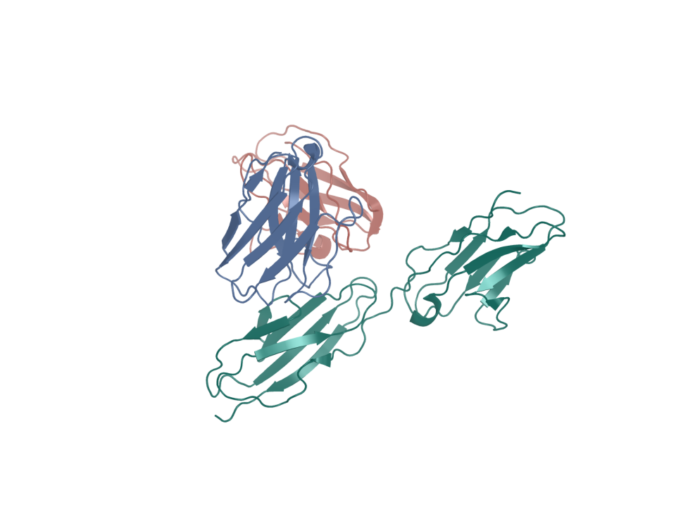
This structure shows the antibody fragment tusamitamab Fab bound to part of CEACAM5, a cell-surface protein.
Open the 3D view to rotate the complex and examine the contact site.
Codex + Rosalind Workbench
Continue the research in Codex
Choose a target or result
Use Target connections or Compare targets to choose a protein. Open its sources, binding sites, and related molecules.
Inspect the molecule
Open Structure records. For a figure with View in 3D, activate the preview, drag to rotate, and scroll to zoom. Use the part controls to isolate chains and label residues.
Continue in Codex
Where a structure offers Copy Codex request, copy it into your task. The request identifies the coordinate file, molecular parts, and a matching sequence when available.
Use the molecular and sequence viewers in Codex to inspect downloaded files. Coordinate and FASTA links appear beside each available structure or sequence.