 Surface Atlas
Explore molecular structures
Rotate a molecular structure, isolate its binding partners, and select residues at the contact site. Use the source links and coordinates to continue your research.
Compare published small-molecule binding with screening poses →
Browse all molecular views
Search published structures, predicted target models, and campaign candidates. Each entry opens its recorded coordinates and links to the source section.
Colors identify molecular parts. Each structure record identifies the construct, chain and residue numbering, and coordinate-file hash.
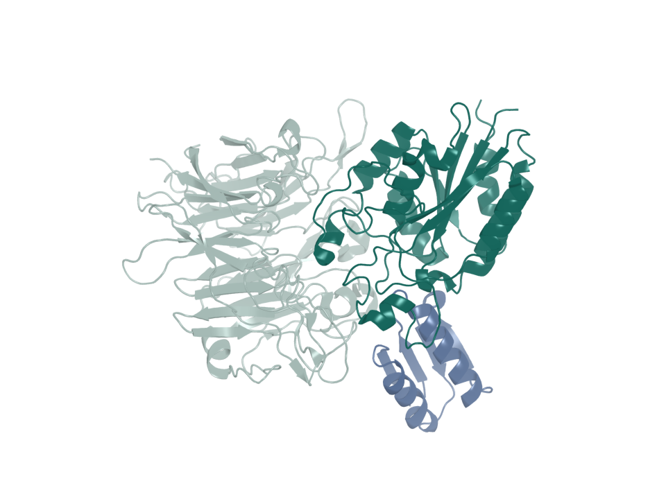
Integrin αvβ6 · published minibinder
B6_BP_dslf is the published reference binder from Roy et al. (2023), developed at the University of Washington Institute for Protein Design with collaborators. The campaign generated separate candidates.
Show the binder alone, then restore the complex to inspect its position.
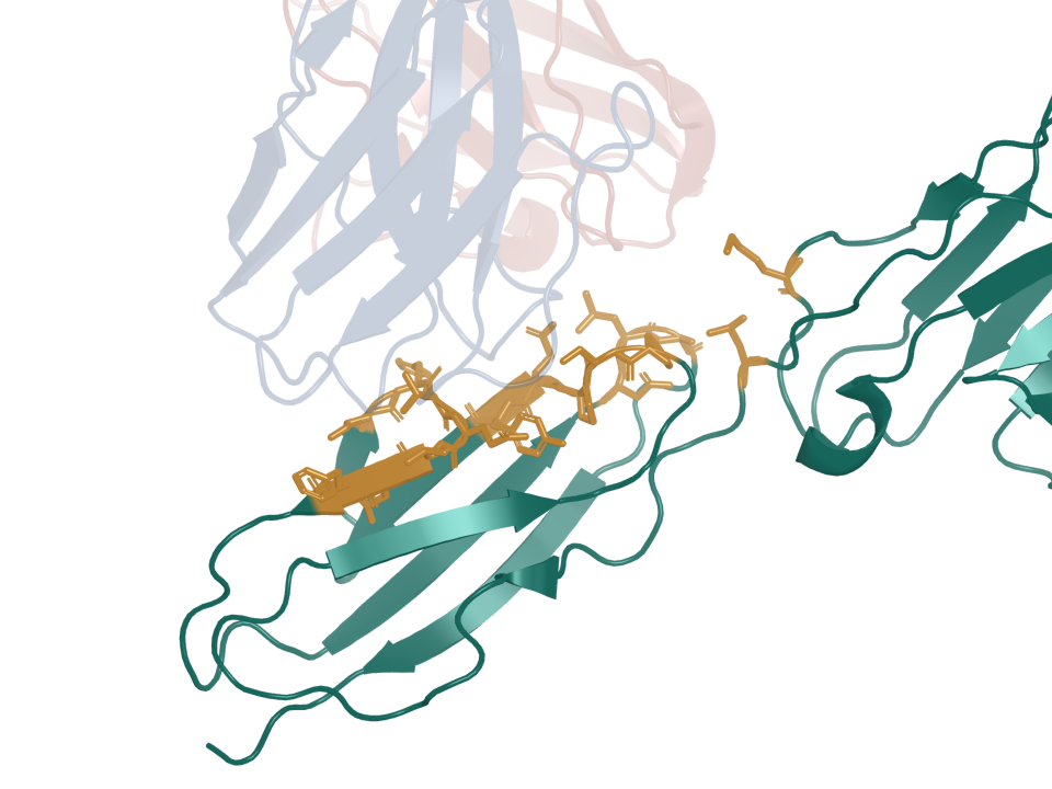
CEACAM5 · antibody contact
This structure shows tusamitamab Fab bound to the CEACAM5 A3–B3 fragment.
Inspect the antibody and target contact residues. The labels retain the deposited chain IDs.
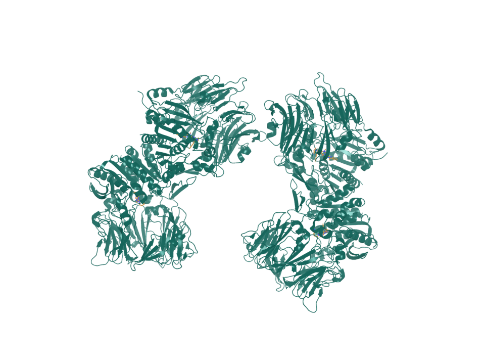
FAP · small-molecule pocket
Linagliptin is the deposited ligand in this FAP reference. Its structure supplies a reference pocket for the computational screen.
Isolate the ligand, then inspect the surrounding target. Read the screening controls before interpreting poses.
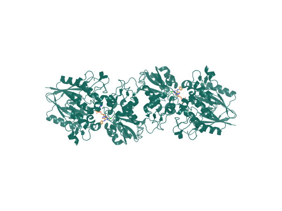
CD73 · small-molecule reference
CD73 is encoded by NT5E. Compound 21 supplies the deposited reference ligand for this screening site.
Compare the molecular partners, then follow the screen to its repeat-run checks.
Compare references with generated candidates
Compare published binding interfaces with generated candidate structures, then inspect each campaign’s sequences and control results.