 Surface Atlas
CEACAM5
CEACAM5 is a cell adhesion protein attached to the cell surface by a lipid anchor.
Protein-binder design
Three methods generated nine CEACAM5 proteins. Reference controls failed in the later prediction study, which stopped candidate selection.
View campaign resultsRNA expression in tumor samples
Median CEACAM5 RNA expression by tumor type, measured in RSEM units. The study selected these samples using microsatellite-stability score thresholds.
Inspect the molecular structure
Published reference structure
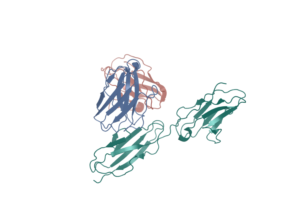
CEACAM5 with tusamitamab
- PDB entry
- 8BW0
- Target ID
- T-ENSG00000105388
- tusamitamab Fab heavy chain · chain H (ribbon)
- tusamitamab Fab light chain · chain L (ribbon)
- CEACAM5 A3-B3 domains · chain C (ribbon)
Sequence & binding sites
Choose a protein region, search amino-acid letters, or inspect a recorded binding site.
Loading sequence…
Open the sequence explorerSequence annotations and source records
Canonical sequence and topology
- Target ID
- T-ENSG00000105388
- UniProt accession
- P06731
- Sequence state
- available
- Canonical sequence
- Yes
- Length in amino acids
- 702
- Sequence SHA-256
- 1d589a5cfe8738ca20f7b2a4658dceebfb6741b489707df585481e190c2d462c
View and select the full canonical sequence
Gray shows the full canonical sequence. Colored spans mark the listed intervals.
The extracellular interval is derived from the signal peptide and GPI attachment site. It is a proposed construct interval.
| Start | End | Length | Sequence and source |
|---|---|---|---|
| 35 | 675 | 641 | Sequence and source
|
Sequence and topology sources
- Source role
- canonical-sequence
- Source
- alphafold-db-canonical-uniprot-field
- Extracted field
- uniprotSequence
- Source row accession
- Not recorded
- Source role
- reviewed-topology
- Source
- UniProt reviewed human surface-field snapshot
- Extracted field
- Not recorded
- Source row accession
- P06731
Download source snapshot (gzip)
- subcellular_location
- SUBCELLULAR LOCATION: Cell membrane {ECO:0000269|PubMed:10864933, ECO:0000269|PubMed:18086185, ECO:0000269|PubMed:2317824, ECO:0000269|PubMed:2803308, ECO:0000269|PubMed:3387431}; Lipid-anchor, GPI-anchor {ECO:0000269|PubMed:10864933, ECO:0000269|PubMed:18086185, ECO:0000269|PubMed:2317824, ECO:0000269|PubMed:2803308, ECO:0000269|PubMed:3387431}; Extracellular side {ECO:0000269|PubMed:10864933, ECO:0000269|PubMed:18086185, ECO:0000269|PubMed:2317824, ECO:0000269|PubMed:2803308, ECO:0000269|PubMed:3387431}. Apical cell membrane {ECO:0000269|PubMed:10436421}. Cell surface {ECO:0000269|PubMed:2317824, ECO:0000269|PubMed:2803308}. Note=Localized to the apical glycocalyx surface. {ECO:0000269|PubMed:10436421}.
- transmembrane
- Not recorded
- topological_domain
- Not recorded
- signal_peptide
- SIGNAL 1..34
- lipidation
- LIPID 676; /note="GPI-anchor amidated alanine"; /evidence="ECO:0000269|PubMed:3387431"
Protein identity and location
- Protein
- Cell adhesion molecule CEACAM5
- Protein location
- GPI-anchored membrane protein
- Canonical sequence
- P06731 · 702 amino acids
- Extracellular region
- An extracellular span at residues 35–675 is derived from the signal peptide and GPI attachment annotation.
More structures and generated models
Structure snapshots
Published reference structure
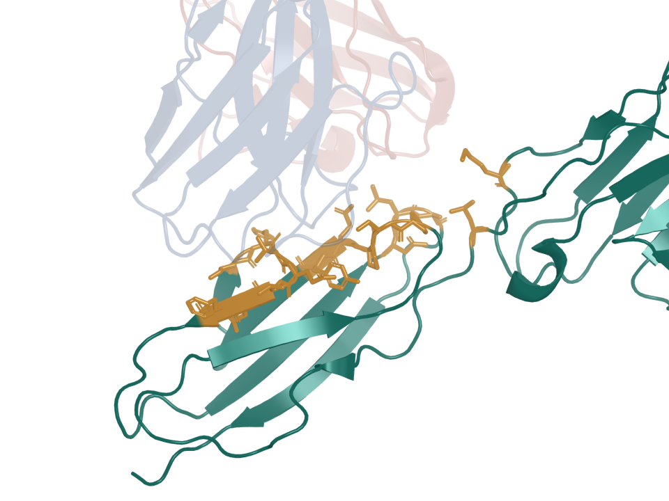
Where tusamitamab contacts CEACAM5
- PDB entry
- 8BW0
- Target ID
- T-ENSG00000105388
- tusamitamab Fab heavy chain · chain H (ribbon)
- tusamitamab Fab light chain · chain L (ribbon)
- CEACAM5 A3-B3 domains · chain C (ribbon)
- CEACAM5 contact residues (sticks)
Predicted complex
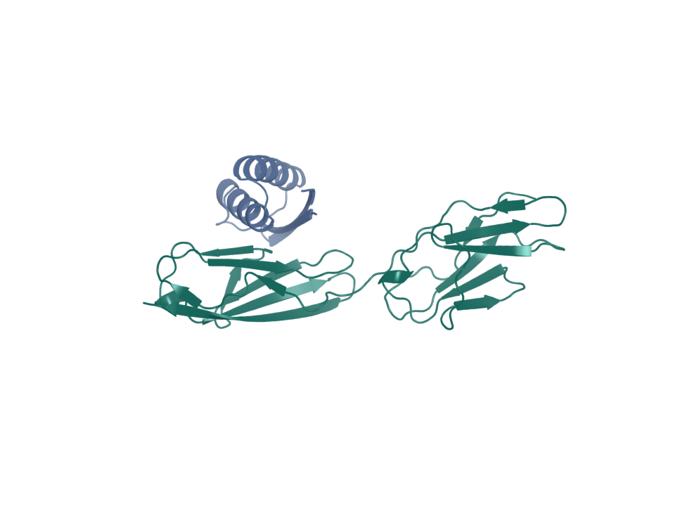
CEACAM5 · BoltzGen 03
- Target ID
- T-ENSG00000105388
- CEACAM5 A3-B3 target construct, 177 residues · chain A (ribbon)
- Designed candidate ceacam5-cp3-boltzgen-03 · chain B (ribbon)
Predicted complex
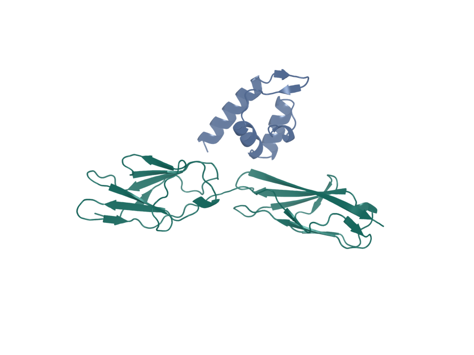
CEACAM5 · Proteina–Complexa 02
- Target ID
- T-ENSG00000105388
- CEACAM5 A3-B3 target construct, 177 residues · chain A (ribbon)
- Designed candidate ceacam5-cp3-proteina-complexa-02 · chain B (ribbon)
Predicted complex
CEACAM5 · Proteina–Complexa 03
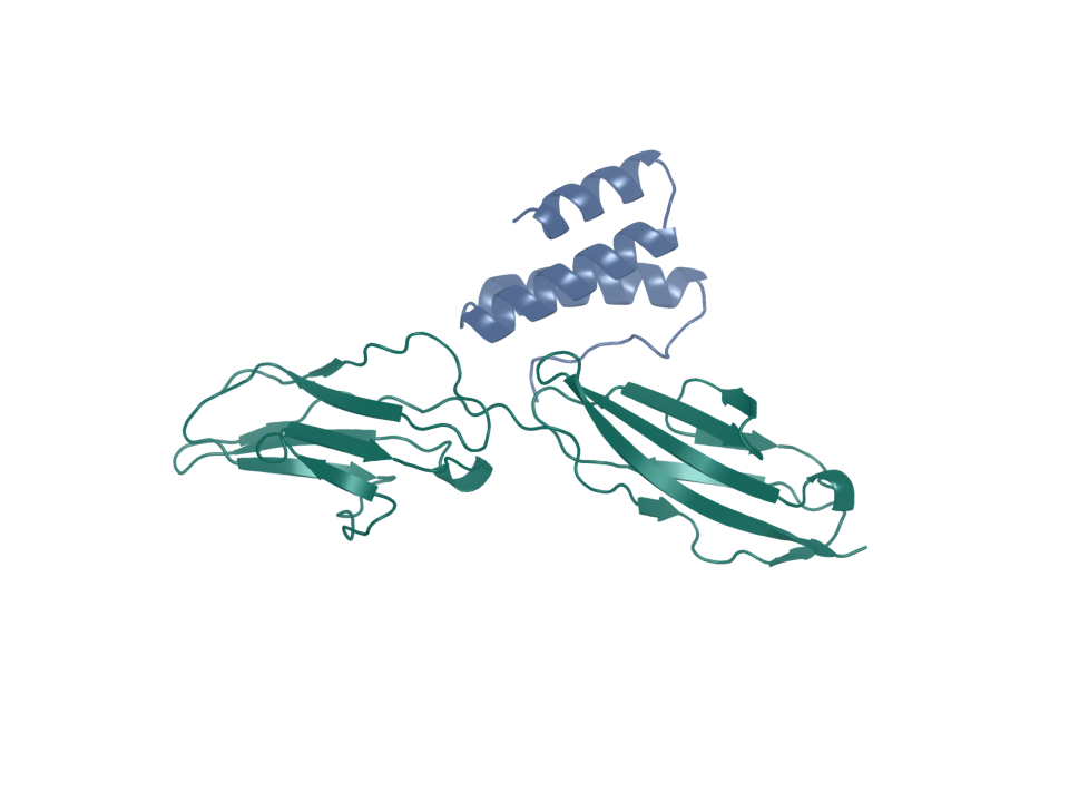
- Target ID
- T-ENSG00000105388
- CEACAM5 A3-B3 target construct, 177 residues · chain A (ribbon)
- Designed candidate ceacam5-cp3-proteina-complexa-03 · chain B (ribbon)
Predicted complex
CEACAM5 · PXDesign–ProteinMPNN 03
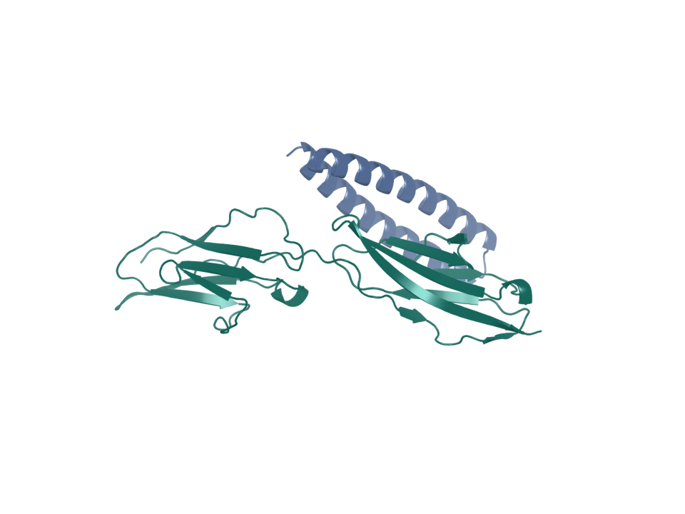
- Target ID
- T-ENSG00000105388
- CEACAM5 A3-B3 target construct, 177 residues · chain A (ribbon)
- Designed candidate ceacam5-cp3-pxdesign-proteinmpnn-03 · chain B (ribbon)
Predicted complex
CEACAM5 · shuffled VH control
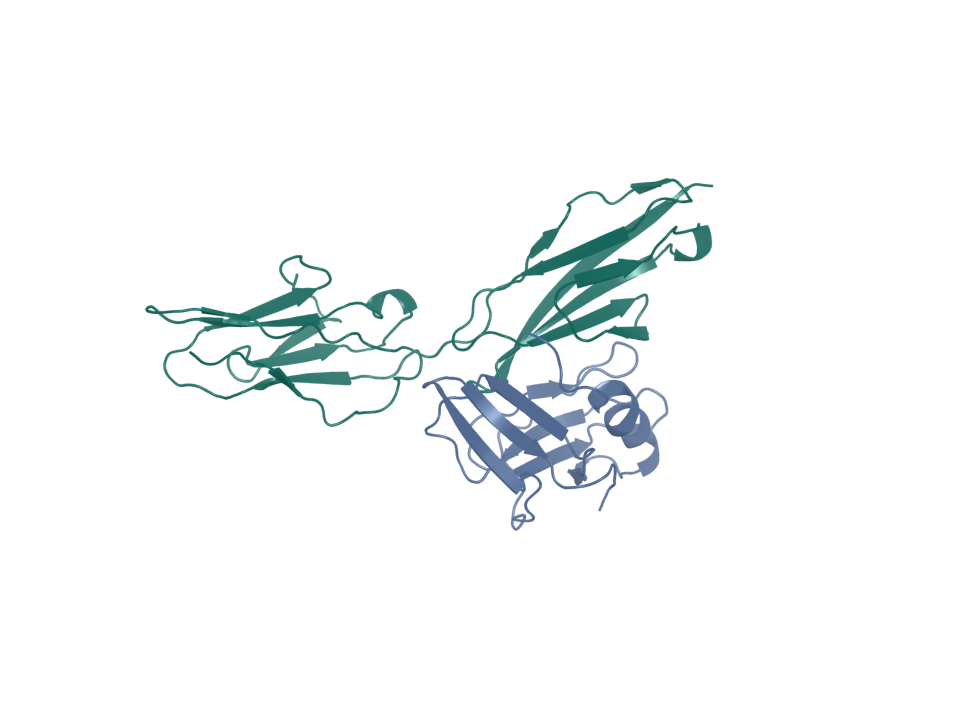
- Target ID
- T-ENSG00000105388
- CEACAM5 A3-B3 target construct, 177 residues · chain A (ribbon)
- Composition-shuffled VH negative control · chain B (ribbon)
Predicted complex
CEACAM5 · paired VH/VL control
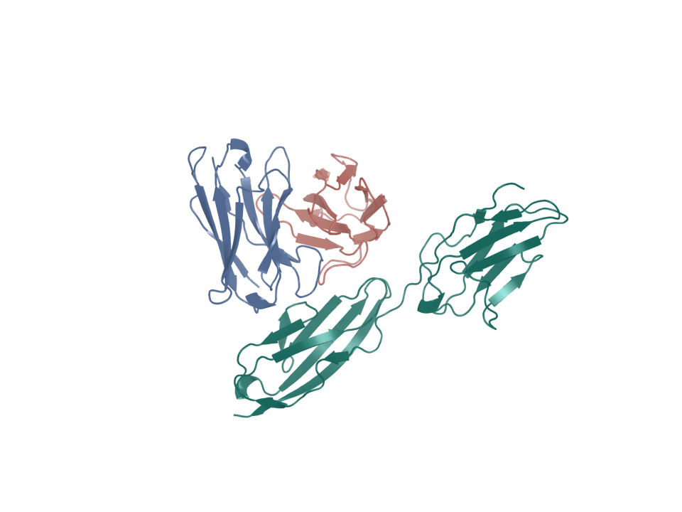
- Target ID
- T-ENSG00000105388
- CEACAM5 target · chain A (ribbon)
- Heavy-chain variable domain (VH) · chain B (ribbon)
- Light-chain variable domain (VL) · chain C (ribbon)
Predicted complex
CEACAM5 · tusamitamab VH control
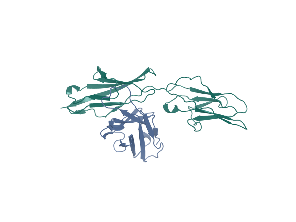
- Target ID
- T-ENSG00000105388
- CEACAM5 A3-B3 target construct, 177 residues · chain A (ribbon)
- Tusamitamab VH positive control · chain B (ribbon)
Expression in normal tissues
- RNA specificity
- Tissue enriched
- RNA distribution
- Detected in many
- RNA tissue-specific nTPM
- intestine: 920.3
- Protein specificity
- Group enriched
- Protein distribution
- Detected in many
- Immunohistochemistry reliability
- Enhanced
- Immunofluorescence reliability
- Supported
- Protein tissue-specific intensity
- intestine: 49,432,922.2; skin: 22,804,857.5
- Tissue cell-type enrichment
- Colon - Colon enterocytes, Minor Salivary Gland - Minor salivary gland basal cells, Prostate - Urothelial cells, Skin - Eccrine sweat gland cells, Stomach - Gastric mucous cells, Thyroid gland - Parafollicular cells
- Subcellular location
- Plasma membrane
Structures
| Name | Stable ID | Action | Format | Status | Claim |
|---|---|---|---|---|---|
| 8BW0 | S-RCSB-8BW0 | Not recorded | Not recorded | not recorded | verified deposited experimental structure |
Sources and downloads
CEACAM5 · ENSG00000105388 · P06731