 Surface Atlas
ERBB2
Receptor tyrosine-protein kinase erbB-2. UniProt annotates extracellular residues 23–652.
Published binding interface
Structure 1N8Z records ERBB2 in contact with trastuzumab Fab. Select the contact residues to inspect this interface.
Open this viewRNA expression by cell type
Percentage of cells with detected ERBB2 RNA in the public colon adenocarcinoma cell groups.
Tumor cells21.7%
Fibroblasts12.6%
Adipocytes6.6%
Endothelial cells3.7%
Dendritic cells3.3%
Natural killer cells2.9%
Macrophages (group B)1.8%
Granulocytes1.4%
CD8 T cells1.0%
CD4 T cells0.8%
Macrophages (group A)0.8%
B cells0.8%
Regulatory T cells0.7%
Inspect the molecular structure
Published reference structure
ERBB2 with trastuzumab
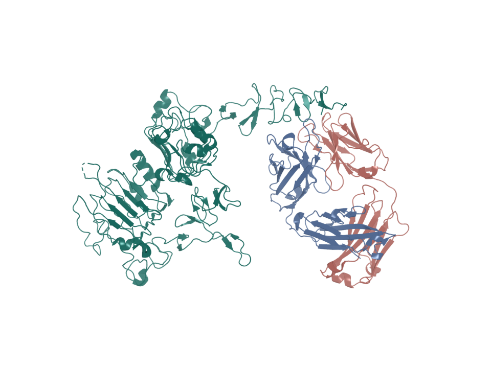
- PDB entry
- 1N8Z
- Target ID
- T-ENSG00000141736
- trastuzumab Fab light chain · chain A (ribbon)
- trastuzumab Fab heavy chain · chain B (ribbon)
- ERBB2 extracellular domain · chain C (ribbon)
Sequence & binding sites
Choose a protein region, search amino-acid letters, or inspect a recorded binding site.
Loading sequence…
Open the sequence explorerSequence annotations and source records
Canonical sequence and topology
- Target ID
- T-ENSG00000141736
- UniProt accession
- P04626
- Sequence state
- available
- Canonical sequence
- Yes
- Length in amino acids
- 1255
- Sequence SHA-256
- a5f256fc653515d110f6dd48ad55b8ea21bf6d45af33b7f5c182c6c31455b636
View and select the full canonical sequence
Gray shows the full canonical sequence. Colored spans mark the listed intervals.
Signal peptide
1–22
Membrane-spanning segments
653–675 (Helical)
Source-annotated domains
23–652 (Extracellular); 676–1255 (Cytoplasmic)
Source-annotated extracellular intervals
23–652
UniProt annotates the following extracellular intervals.
| Start | End | Length | Sequence and source |
|---|---|---|---|
| 23 | 652 | 630 | Sequence and source
|
Sequence and topology sources
- Source role
- canonical-sequence
- Source
- alphafold-db-canonical-uniprot-field
- Extracted field
- uniprotSequence
- Source row accession
- Not recorded
- Source role
- reviewed-topology
- Source
- UniProt reviewed human surface-field snapshot
- Extracted field
- Not recorded
- Source row accession
- P04626
Download source snapshot (gzip)
- subcellular_location
- SUBCELLULAR LOCATION: Cell membrane {ECO:0000269|PubMed:32381043}; Single-pass type I membrane protein {ECO:0000255}. Cell projection, ruffle membrane {ECO:0000269|PubMed:34380438}; Single-pass type I membrane protein {ECO:0000255}. Note=Internalized from the cell membrane in response to EGF stimulation. {ECO:0000269|PubMed:32381043}.; SUBCELLULAR LOCATION: [Isoform 1]: Cell membrane {ECO:0000269|PubMed:31138794, ECO:0000269|PubMed:33497358}; Single-pass type I membrane protein {ECO:0000255}. Early endosome {ECO:0000269|PubMed:31138794}. Cytoplasm, perinuclear region. Nucleus. Note=Translocation to the nucleus requires endocytosis, probably endosomal sorting and is mediated by importin beta-1/KPNB1. Also detected in VPS35-positive endosome-to-TGN retrograde vesicles (PubMed:31138794). {ECO:0000269|PubMed:31138794}.; SUBCELLULAR LOCATION: [Isoform 2]: Cytoplasm. Nucleus.; SUBCELLULAR LOCATION: [Isoform 3]: Cytoplasm. Nucleus.
- transmembrane
- TRANSMEM 653..675; /note="Helical"; /evidence="ECO:0000255"
- topological_domain
- TOPO_DOM 23..652; /note="Extracellular"; /evidence="ECO:0000255"; TOPO_DOM 676..1255; /note="Cytoplasmic"; /evidence="ECO:0000255"
- signal_peptide
- SIGNAL 1..22; /evidence="ECO:0000255"
- lipidation
- Not recorded
Protein identity and location
- Protein
- Receptor tyrosine-protein kinase erbB-2
- Protein location
- Transmembrane protein
- Canonical sequence
- P04626 · 1,255 amino acids
- Extracellular region
- UniProt annotates extracellular residues 23–652.
More structures and generated models
Structure snapshots
Expression in normal tissues
- RNA specificity
- Low tissue specificity
- RNA distribution
- Detected in all
- Protein specificity
- Tissue enriched
- Protein distribution
- Detected in many
- Immunohistochemistry reliability
- Enhanced
- Immunofluorescence reliability
- Enhanced
- Protein tissue-specific intensity
- lymphoid tissue: 8,296,603.9
- Tissue cell-type enrichment
- Adrenal gland - Fibroblasts, Breast - Breast glandular cells, Lung - Alveolar cells type 1, Pituitary gland - Undifferentiated cells (Pituitary gland), Spleen - Fibroblasts_1
- Subcellular location
- Plasma membrane
Structures
| Name | Stable ID | Action | Format | Status | Claim |
|---|---|---|---|---|---|
| 1N8Z | S-RCSB-1N8Z | Not recorded | Not recorded | not recorded | verified deposited experimental structure |
Sources and downloads
ERBB2 · ENSG00000141736 · P04626