 Surface Atlas
FAP
Prolyl endopeptidase FAP. UniProt annotates extracellular residues 26–760.
Molecule screening
The FAP campaign screened 24 molecules and repeated five selections. All five passed the recorded docking and pose checks.
View campaign resultsRNA expression by cell type
Percentage of cells with detected FAP RNA in the public colon adenocarcinoma cell groups.
Inspect the molecular structure
Published reference structure
FAP with linagliptin
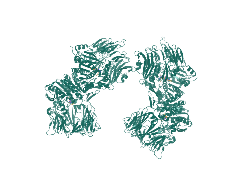
- PDB entry
- 6Y0F
- Target ID
- T-ENSG00000078098
- FAP · chains A, B, C, D (ribbon)
- Linagliptin · carbon in ochre; oxygen red; nitrogen blue (sticks)
Sequence & binding sites
Choose a protein region, search amino-acid letters, or inspect a recorded binding site.
Loading sequence…
Open the sequence explorerSequence annotations and source records
Canonical sequence and topology
- Target ID
- T-ENSG00000078098
- UniProt accession
- Q12884
- Sequence state
- available
- Canonical sequence
- Yes
- Length in amino acids
- 760
- Sequence SHA-256
- fc502b7363283bca3ea0e276ed1d65d0ae8d5064519a226a06f9c77059235b6f
View and select the full canonical sequence
Gray shows the full canonical sequence. Colored spans mark the listed intervals.
UniProt annotates the following extracellular intervals.
| Start | End | Length | Sequence and source |
|---|---|---|---|
| 26 | 760 | 735 | Sequence and source
|
Sequence and topology sources
- Source role
- canonical-sequence
- Source
- alphafold-db-canonical-uniprot-field
- Extracted field
- uniprotSequence
- Source row accession
- Not recorded
- Source role
- reviewed-topology
- Source
- UniProt reviewed human surface-field snapshot
- Extracted field
- Not recorded
- Source row accession
- Q12884
Download source snapshot (gzip)
- subcellular_location
- SUBCELLULAR LOCATION: [Prolyl endopeptidase FAP]: Cell surface {ECO:0000269|PubMed:10593948, ECO:0000269|PubMed:16175601, ECO:0000269|PubMed:17105646, ECO:0000269|PubMed:24717288, ECO:0000269|PubMed:7911242}. Cell membrane {ECO:0000269|PubMed:12376466, ECO:0000269|PubMed:16651416, ECO:0000269|PubMed:9065413, ECO:0000303|PubMed:10455171}; Single-pass type II membrane protein {ECO:0000255}. Cell projection, lamellipodium membrane {ECO:0000269|PubMed:16651416, ECO:0000269|PubMed:9065413}; Single-pass type II membrane protein {ECO:0000255}. Cell projection, invadopodium membrane {ECO:0000269|PubMed:12376466, ECO:0000269|PubMed:16651416, ECO:0000269|PubMed:7923219, ECO:0000269|PubMed:9065413, ECO:0000303|PubMed:10455171}; Single-pass type II membrane protein {ECO:0000255}. Cell projection, ruffle membrane {ECO:0000303|PubMed:10455171}; Single-pass type II membrane protein {ECO:0000255}. Membrane {ECO:0000269|PubMed:2172980}; Single-pass type II membrane protein {ECO:0000255}. Note=Localized on cell surface with lamellipodia and invadopodia membranes and on shed vesicles. Colocalized with DPP4 at invadopodia and lamellipodia membranes of migratory activated endothelial cells in collagenous matrix. Colocalized with DPP4 on endothelial cells of capillary-like microvessels but not large vessels within invasive breast ductal carcinoma. Anchored and enriched preferentially by integrin alpha-3/beta-1 at invadopodia, plasma membrane protrusions that correspond to sites of cell invasion, in a collagen-dependent manner. Localized at plasma and ruffle membranes in a collagen-independent manner. Colocalized with PLAUR preferentially at the cell surface of invadopodia membranes in a cytoskeleton-, integrin- and vitronectin-dependent manner. Concentrated at invadopodia membranes, specialized protrusions of the ventral plasma membrane in a fibrobectin-dependent manner. Colocalizes with extracellular components (ECM), such as collagen fibers and fibronectin. {ECO:0000269|PubMed:10593948, ECO:0000269|PubMed:12376466, ECO:0000269|PubMed:16175601, ECO:0000269|PubMed:16651416, ECO:0000269|PubMed:17105646, ECO:0000269|PubMed:2172980, ECO:0000269|PubMed:24717288, ECO:0000269|PubMed:7911242, ECO:0000269|PubMed:7923219, ECO:0000269|PubMed:9065413, ECO:0000303|PubMed:10455171}.; SUBCELLULAR LOCATION: [Antiplasmin-cleaving enzyme FAP, soluble form]: Secreted {ECO:0000269|PubMed:14751930, ECO:0000269|PubMed:16223769, ECO:0000269|PubMed:24371721}. Note=Found in blood plasma and serum. {ECO:0000269|PubMed:14751930, ECO:0000269|PubMed:16223769, ECO:0000269|PubMed:24371721}.; SUBCELLULAR LOCATION: [Isoform 2]: Cytoplasm {ECO:0000303|PubMed:10644713}.
- transmembrane
- TRANSMEM 5..25; /note="Helical; Signal-anchor for type II membrane protein"; /evidence="ECO:0000255"
- topological_domain
- TOPO_DOM 1..4; /note="Cytoplasmic"; /evidence="ECO:0000255, ECO:0000303|PubMed:14751930"; TOPO_DOM 26..760; /note="Extracellular"; /evidence="ECO:0000255, ECO:0000303|PubMed:14751930"
- signal_peptide
- Not recorded
- lipidation
- Not recorded
Protein identity and location
- Protein
- Prolyl endopeptidase FAP
- Protein location
- Transmembrane protein
- Canonical sequence
- Q12884 · 760 amino acids
- Extracellular region
- UniProt annotates extracellular residues 26–760.
More structures and generated models
Structure snapshots
Published reference structure
FAP with a SUMO-I3 VHH fusion
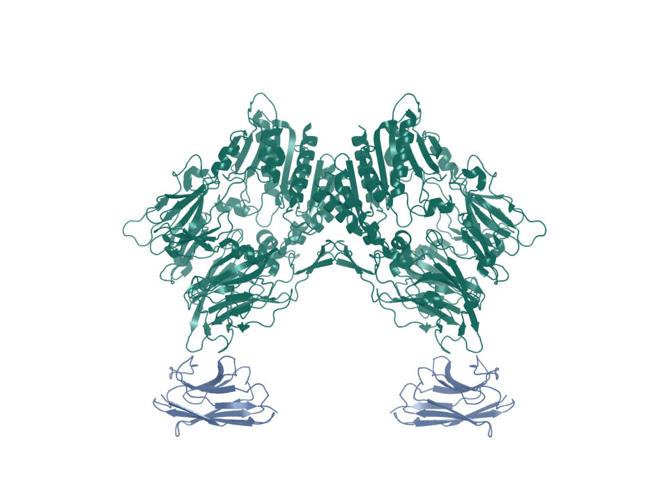
- PDB entry
- 9DVR
- Target ID
- T-ENSG00000078098
- FAP soluble ectodomain dimer · chains A, B (ribbon)
- SUMO-I3 VHH fusion · chains G, H (ribbon)
Expression in normal tissues
- RNA specificity
- Tissue enhanced
- RNA distribution
- Detected in many
- RNA tissue-specific nTPM
- endometrium 1: 29.4
- Protein specificity
- Group enriched
- Protein distribution
- Detected in some
- Immunohistochemistry reliability
- Uncertain
- Protein tissue-specific intensity
- blood vessel: 187,180.7; skin: 538,235.3
- Tissue cell-type enrichment
- Adipose subcutaneous - Adipose progenitor cells, Adipose visceral - Adipose progenitor cells, Minor Salivary Gland - Macrophages, Prostate - Fibroblasts, Skeletal muscle - Fibroblasts, Testis - Early spermatids, Thyroid gland - Fibroblasts
Known molecule interactions
| Molecule | Stable ID | Category | Reference or intention | Claim |
|---|---|---|---|---|
| Linagliptin | RF020 | Reference ligand | source | source-backed known interaction |
Molecule screening results
The FAP campaign screened 24 molecules and repeated five selections. All five passed the recorded docking and pose checks.
Compare molecules, poses, and repeated runs →Structures
| Name | Stable ID | Action | Format | Status | Claim |
|---|---|---|---|---|---|
| 9DVR | S-RCSB-9DVR | Not recorded | Not recorded | not recorded | verified deposited experimental structure |
| 6Y0F | S-RCSB-6Y0F | Not recorded | Not recorded | not recorded | verified deposited experimental structure |
Sources and downloads
FAP · ENSG00000078098 · Q12884