 Surface Atlas
ITGAV
Integrin alpha-V. UniProt annotates extracellular residues 31–992.
Published binding interface
Structure 8TCG records ITGAV in contact with B6_BP_dslf miniprotein. Select the contact residues to inspect this interface.
Open this viewRNA expression by cell type
Percentage of cells with detected ITGAV RNA in the public colon adenocarcinoma cell groups.
Fibroblasts39.4%
Endothelial cells29.1%
Macrophages (group B)22.9%
Adipocytes16.2%
Tumor cells12.6%
Macrophages (group A)7.3%
Dendritic cells5.9%
Granulocytes4.6%
Regulatory T cells3.4%
Natural killer cells2.4%
B cells1.8%
CD8 T cells1.7%
CD4 T cells1.7%
Inspect the molecular structure
Published reference structure
Integrin alpha-v beta-6 with a minibinder
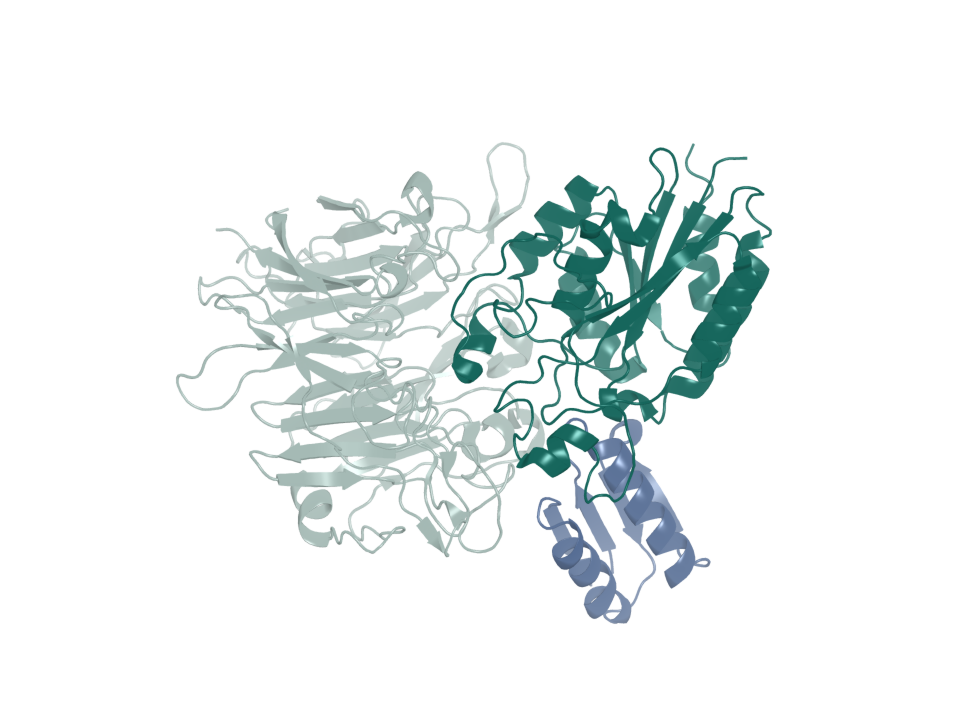
- PDB entry
- 8TCG
- Target ID
- T-ENSG00000138448
- integrin alpha-V · chain A (ribbon)
- integrin beta-6 · chain B (ribbon)
- B6_BP_dslf minibinder · chain C (ribbon)
Published minibinder: Roy et al., 2023 · PDB 8TCG
Sequence & binding sites
Choose a protein region, search amino-acid letters, or inspect a recorded binding site.
Loading sequence…
Open the sequence explorerSequence annotations and source records
Canonical sequence and topology
- Target ID
- T-ENSG00000138448
- UniProt accession
- P06756
- Sequence state
- available
- Canonical sequence
- Yes
- Length in amino acids
- 1048
- Sequence SHA-256
- d5e1e101edb8e81ca39900444877a2dcc9e6080adffde94a47d58b0ee6f6c811
View and select the full canonical sequence
Gray shows the full canonical sequence. Colored spans mark the listed intervals.
Signal peptide
1–30
Membrane-spanning segments
993–1016 (Helical)
Source-annotated domains
31–992 (Extracellular); 1017–1048 (Cytoplasmic)
Source-annotated extracellular intervals
31–992
UniProt annotates the following extracellular intervals.
| Start | End | Length | Sequence and source |
|---|---|---|---|
| 31 | 992 | 962 | Sequence and source
|
Sequence and topology sources
- Source role
- canonical-sequence
- Source
- alphafold-db-canonical-uniprot-field
- Extracted field
- uniprotSequence
- Source row accession
- Not recorded
- Source role
- reviewed-topology
- Source
- UniProt reviewed human surface-field snapshot
- Extracted field
- Not recorded
- Source row accession
- P06756
Download source snapshot (gzip)
- subcellular_location
- SUBCELLULAR LOCATION: Cell membrane; Single-pass type I membrane protein. Cell junction, focal adhesion {ECO:0000269|PubMed:17158881}.
- transmembrane
- TRANSMEM 993..1016; /note="Helical"; /evidence="ECO:0000255"
- topological_domain
- TOPO_DOM 31..992; /note="Extracellular"; /evidence="ECO:0000255"; TOPO_DOM 1017..1048; /note="Cytoplasmic"; /evidence="ECO:0000255"
- signal_peptide
- SIGNAL 1..30; /evidence="ECO:0000269|PubMed:2467745"
- lipidation
- Not recorded
Protein identity and location
- Protein
- Integrin alpha-V
- Protein location
- Transmembrane protein
- Canonical sequence
- P06756 · 1,048 amino acids
- Extracellular region
- UniProt annotates extracellular residues 31–992.
More structures and generated models
Structure snapshots
Expression in normal tissues
- RNA specificity
- Low tissue specificity
- RNA distribution
- Detected in all
- Protein specificity
- Tissue enhanced
- Protein distribution
- Detected in all
- Immunohistochemistry reliability
- Supported
- Immunofluorescence reliability
- Approved
- Protein tissue-specific intensity
- blood vessel: 145,026,787.2
- Tissue cell-type enrichment
- Adrenal gland - Adrenal cortex cells
- Subcellular location
- Focal adhesion sites, Cytosol
Structures
| Name | Stable ID | Action | Format | Status | Claim |
|---|---|---|---|---|---|
| 8TCG | S-RCSB-8TCG | Not recorded | Not recorded | not recorded | verified deposited experimental structure |
| 8TCG | S-RCSB-8TCG-RFD3-AB | Not recorded | Not recorded | not recorded | hash-bound target preparation and observed deposited interface geometry |
Sources and downloads
ITGAV · ENSG00000138448 · P06756