 Surface Atlas
ITGB6
Integrin beta-6. UniProt annotates extracellular residues 22–709.
Protein-binder design
The campaign generated 16 binder designs with sequences and 3D models. None of the 14 scored complexes met the binding-site geometry criteria.
View campaign resultsRNA expression by cell type
Percentage of cells with detected ITGB6 RNA in the public colon adenocarcinoma cell groups.
Tumor cells8.5%
Fibroblasts1.0%
Endothelial cells0.5%
Natural killer cells0.5%
Macrophages (group B)0.4%
Adipocytes0.3%
Macrophages (group A)0.3%
CD8 T cells0.2%
CD4 T cells0.2%
Regulatory T cells0.2%
B cells0.1%
Dendritic cells0.0%
Granulocytes0.0%
Inspect the molecular structure
Prepared target
Integrin alpha-v beta-6: prepared target
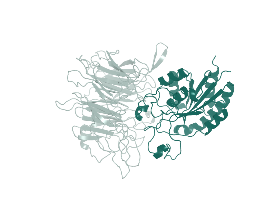
- PDB entry
- 8TCG
- Target ID
- T-ENSG00000115221
- Integrin alpha-v · chain A (ribbon)
- Integrin beta-6 · chain B (ribbon)
Sequence & binding sites
Choose a protein region, search amino-acid letters, or inspect a recorded binding site.
Loading sequence…
Open the sequence explorerSequence annotations and source records
Canonical sequence and topology
- Target ID
- T-ENSG00000115221
- UniProt accession
- P18564
- Sequence state
- available
- Canonical sequence
- Yes
- Length in amino acids
- 788
- Sequence SHA-256
- 871c82602ef83b96fc7e48abb1d783377410233b25a6bf2f2850d98965d21860
View and select the full canonical sequence
Gray shows the full canonical sequence. Colored spans mark the listed intervals.
Signal peptide
1–21
Membrane-spanning segments
710–730 (Helical)
Source-annotated domains
22–709 (Extracellular); 731–788 (Cytoplasmic)
Source-annotated extracellular intervals
22–709
UniProt annotates the following extracellular intervals.
| Start | End | Length | Sequence and source |
|---|---|---|---|
| 22 | 709 | 688 | Sequence and source
|
Sequence and topology sources
- Source role
- canonical-sequence
- Source
- alphafold-db-canonical-uniprot-field
- Extracted field
- uniprotSequence
- Source row accession
- Not recorded
- Source role
- reviewed-topology
- Source
- UniProt reviewed human surface-field snapshot
- Extracted field
- Not recorded
- Source row accession
- P18564
Download source snapshot (gzip)
- subcellular_location
- SUBCELLULAR LOCATION: Cell membrane {ECO:0000269|PubMed:17545607}; Single-pass type I membrane protein {ECO:0000305}. Cell junction, focal adhesion {ECO:0000269|PubMed:17158881}.
- transmembrane
- TRANSMEM 710..730; /note="Helical"; /evidence="ECO:0000255"
- topological_domain
- TOPO_DOM 22..709; /note="Extracellular"; /evidence="ECO:0000255"; TOPO_DOM 731..788; /note="Cytoplasmic"; /evidence="ECO:0000255"
- signal_peptide
- SIGNAL 1..21; /evidence="ECO:0000255"
- lipidation
- Not recorded
Protein identity and location
- Protein
- Integrin beta-6
- Protein location
- Transmembrane protein
- Canonical sequence
- P18564 · 788 amino acids
- Extracellular region
- UniProt annotates extracellular residues 22–709.
More structures and generated models
Structure snapshots
Expression in normal tissues
- RNA specificity
- Tissue enhanced
- RNA distribution
- Detected in many
- RNA tissue-specific nTPM
- skeletal muscle: 74.9; tongue: 60.3
- Protein specificity
- Low tissue specificity
- Protein distribution
- Detected in many
- Immunohistochemistry reliability
- Approved
- Immunofluorescence reliability
- Approved
- Tissue cell-type enrichment
- Colon - Colon enterocytes, Lung - Alveolar cells type 2, Pancreas - Ductal cells, Prostate - Urothelial cells, Skeletal muscle - Skeletal myocytes, Stomach - Gastric mucous cells
- Subcellular location
- Nucleoplasm, Cell Junctions, Acrosome
Structures
| Name | Stable ID | Action | Format | Status | Claim |
|---|---|---|---|---|---|
| 8TCG | S-RCSB-8TCG | Not recorded | Not recorded | not recorded | verified deposited experimental structure |
| 8TCG | S-RCSB-8TCG-RFD3-AB | Not recorded | Not recorded | not recorded | hash-bound target preparation and observed deposited interface geometry |
Sources and downloads
ITGB6 · ENSG00000115221 · P18564